Understanding Breast Implant-Associated Anaplastic Large Cell Lymphoma
A form of T-cell (non-Hodgkin) lymphoma known as breast implant associated anaplastic large-cell lymphoma (BIA-ALCL) has been noted as a rare association with the implant-based approach of reconstructive surgery post mastectomy.
David Leos, RN, MBA, OCN
For patients with breast cancer choosing to undergo reconstructive surgery post mastectomy, the options include either the use of autologous tissue flaps (free or pedicled) or a prosthetic implant—based approach with prior use of tissue expanders (2-stage) or without (1stage).
The complication/risk profile for the implant-based approach includes these possibilities: infection, hematoma/seroma, capsular contraction around the implant, pain, changes in breast sensation, implant rupture/silicone gel leakage, and rheumatologic issues, to name a few.
Relatively recently, a little-known and rare association with a form of T-cell (non-Hodgkin) lymphoma known as breast implant associated anaplastic large-cell lymphoma (BIA-ALCL) has been described. Among the first cases reported were in the mid-1990s.1,2
Primary lymphomas of the breast in and of themselves are uncommon; on the rare occasion that BIA-ALCL develops, it occurs as a primary disease in close proximity to the implant. The incidence of this disease is estimated at 0.1 to 0.3 per 100,000 women with breast implants per year.3 BIA-ALCL commonly presents as a late seroma and/or tumor mass attached to the scar capsule containing malignant cells an average of 5.8 years after implant placement (range, 0.4—20 years).4
Subclassifications of ALCL include: systemic, primary cutaneous, and secondary. While systemic and secondary cases are frequently aggressive and express anaplastic large-cell kinase (ALK+), BIA-ALCL tends to have an indolent clinical course and is usually ALK negative.5
The clinical presentation is largely unilateral and characterized by an accumulation of fluid around the implant referred to as seroma. This occurs most commonly, if not exclusively,6 in textured surface versions of both saline and silicone implants as opposed to the smooth surface variety. One theory is that the non-smooth surface of these implants’ outer linings can create a shearing/abrasive effect within the tissue capsule that over time can trigger inflammation that then stimulates an immune response that may subsequently transform into a lymphoma. Biomarkers also provide evidence that underlying inflammation is the likely initiator of this disease.7
Cases of BIA-ALCL confined to the capsule area are considered early-stage disease and tend to have an indolent clinical course. Treatment options include surgical removal of the affected implant and the fibrous capsule. Locally advanced disease may require chemotherapy and radiation therapy in addition to surgical excision. There is reason to believe that implant-associated ALCL is more similar to primary cutaneous ALCL than to systemic ALCL which could favor a surgical remedy over the need for chemotherapy or radiation.8
Much remains to be learned about this disease to prevent delay in diagnosis and inappropriate treatment.9 Lack of routine pathology is likely to delay the diagnosis when women with implants seek medical care from plastic surgeons or primary care physicians for swollen breasts that are assumed to be infected.10
Guidance for Patients
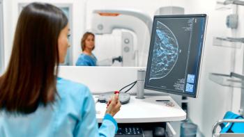
The FDA recommends that all women with silicone gel breast implants undergo regular MRI scans starting 3 years after implantation10 to check for implant rupture, but most insurance policies do not cover such screening; therefore, few women follow these guidelines.11 The agency has also issued a series of safety notices pertaining to data collected thus far. The American Society of Plastic Surgeons and the FDA have jointly established the PROFILE registry to facilitate further determination of an association and is thus set up to capture reports of any confirmed cases.
In a recent article, the presence of biofilm associated with the textured variety of implants appears to have an association with the development of BIA-ALCL.12 This high bacterial load produces a significant and linear increase in lymphocyte activation.13
Research by Sahr et al suggests that gram-negative organisms, in particular Ralstonia pickettii, may stimulate T-cell proliferation which then leads to the development of BIA-ALCL.14
Biofilm is familiar to oncology nurses who work with implanted ports. The presence of this protective buildup of biofilm within the reservoir of the port provides a spawning ground for the bacteria that lead to central line—associated bloodstream infections. Similarly, textured implant surfaces that are meant to stabilize movement offer an anchoring opportunity for bacterial colonization. The bacteria could be exogenously introduced despite conscientious surgical technique during implantation or could be part of the preexisting microbiome of the breast.
Implications of BIA-ALCL and Resources for the Oncology Nurse
Although this condition can evolve from implant use in both the aesthetic augmentation setting as well as that of postmastectomy reconstructive breast surgery, its impact is particularly salient to the oncology nurse.
In the former setting, implants can be in place for decades. For the latter, survivorship of patients with early-stage breast cancer, many of whom have opted for implant-based breast reconstruction after mastectomy, oftentimes spans well over a decade from diagnosis. In either case, there is sufficient time for this condition, albeit rarely, to manifest itself.
Oncology nurses, being familiar with not only lymphomas, but also the concept of secondary malignancy risks related to ablative chemotherapy and/or radiation treatment, are prepared to consider this admittedly low risk in a proper context.
While the pathogenesis of BIA-ALCL is not definitively known at present, discouraging women from undergoing implant surgery is not believed to be warranted. Through awareness, nurses can provide guidance to their patients who’ve survived breast cancer and gone through reconstruction as part of their rehabilitation back to a semblance of their prior norm.
Although not a secondary malignancy in the usual sense, nurses can be prepared to improve awareness of this possibility and the appropriate diagnostic and treatment resources. These include the NCCN guidelines (v2.2017) for initial and pathologic workup, staging, and treatment.15 Fortunately, early-stage disease (localized to the capsule/implant/breast) can be effectively diagnosed via cytology with immunohistochemical and flow cytometry testing for T-cell markers and CD30. Incomplete resection or inadequate local surgical control may subject the patient to recurrence, or to the need for adjunctive treatments such as chemotherapy and radiation therapy, whereas complete resection may be the definitive treatment in the majority of cases.16
A unifying hypothesis to explain this condition is offered by Loch-Wilkenson and colleagues.17 It states that contamination of textured implants with higher surface area by bacteria leading to chronic antigen stimulation in genetically susceptible hosts over a prolonged period of time may result in transformation of T-cells into BIA-ALCL.
References
- Duvic M, Moore D, Menter A, et al. Cutaneous T-cell lymphoma in association with silicone breast implants. J Am Acad Dermatol.1995;32:939-942.
- Keech JA, Creech BJ. Anaplastic T-cell lymphoma in proximity to a saline-filled breast implant. Plast Reconstr Surg. 1997;100:554-555.
- de Jong D, Vasmel WL, de Boer JP, et al. Anaplastic large-cell lymphoma in women with breast implants. JAMA. 2008; 300:2030-2035.
- Stein H, Foss D, Durkop H, et al. CD30(+) anaplastic large cell lymphoma: a review of the histopathologic, genetic, and clinical features. Blood. 2000;96(12):3681-3695.
- Querfeld C, Kuzel TM, Guitart J, Rosen ST. Primary cutaneous CD30+ lymphoproliferative disorders: new insights into biology and therapy. Oncology. 2007;21:689-700.
- Doren EL, Miranda RN, Selber JC, et al. US Epidemiology of Breast Implant-Associated Anaplastic Large Cell Lymphoma. Plast Reconstr Surg. 2017; 139(5):1042-1050.
- Kadin ME, Deva A, Xu H, et al. Biomarkers provide clues to early events in the pathogenesis of breast implant- associated anaplastic large cell lymphoma. Aesthet Surg J. 2016;36:773-781.
- Kim B, Roth C, Chung, KC, Young VL, et al. Anaplastic large cell lymphoma and breast implants: A systematic review. Plast Reconstr Surg. 2011;127:2141-2150.
- Rupani A, Frame JD, Kamel D. Lymphomas associated with breast implants: a review of the literature. Aesthet Surg J.2015;35(5):533-544.
- US Food and Drug Administration. Reports of anaplastic large cell lymphoma in women with breast implants: FDA safety communication. https://www.fda.gov/medicaldevices/productsandmedicalprocedures/implantsandprosthetics/breastimplants/ucm239995.htm. Accessed May 20, 2017.
- Mazzuco AE. Next steps for breast implant—associated anaplastic large-cell lymphoma. Editorial. J Clin Onc2014;21(32):2275.
- Jacombs A, Tahir S, Hu H, et al. In vitro and in vivo investigation of the influence of implant surface on the formation of bacterial biofilm in mammary implants. Plast Reconstr Surg. 2014;133(4):471e-480e.
- Hu H, Johani K, Almatroudi A, et al. Bacterial biofilm infection detected in breast implant—associated anaplastic large-cell lymphoma. Plast. Reconstr Surg. 2016;137(6):1659-1669.
- Sahr A, Former S, Hildebrand D, Heeg K. T-cell activation or tolerization: the yin and yang of bacterial superantigens. Front Microbiol. 2015;6:1153.
- National Comprehensive Cancer Network. Breast Implant Associated ALCL: Guidelines Version 2.2017. https://www.nccn.org/professionals/physician_gls/pdf/t-cell.pdf. Accessed May 20, 2017.
- Clemens MW, Nava MB, Rocco N, Miranda RN. Understanding rare adverse sequelae of breast implants: anaplastic large-cell lymphoma, late seromas, and double capsules. Gland Surg. 2017;6(2):169-184.
- Loch-Wilkenson A, Beath K, Knight RJW, et al. Breast implant associated anaplastic large cell lymphoma in Australia and New Zealand - high surface area textured implants are associated with increased risk [published online ahead of print May 5, 2017]. Plast Reconstr Surg.
David Leos, RN, MBA, OCN, is Manager, Clinical Protocol Administration, with the Department of Plastic Surgery at The University of Texas MD Anderson Cancer Center in Houston, a referral center for these patients.
Newsletter
Knowledge is power. Don’t miss the most recent breakthroughs in cancer care.


